Dr. S. Arumugam has operated on more than 3000 patients in his career and helped change the lives of many more. His team of anesthesiologists and nurses are thoroughly equipped to care for patients with hip conditions.
Some of the most common arthroscopic preocedures that the doctor deals with on a regular basis are:
- Meniscus debridement or repair
- Removal of loose bodies
- Treatment of isolated cartilage defects
- Anterior Cruciate Ligament Reconstruction
- Scar Tissue Releases
Meniscus Debridement
What is a Meniscus?
Meniscus is a soft, C-shaped tissue that lies between the leg bone and the thigh bone. It acts as a shock absorber and helps distribute the body weight evenly across the knee. It decreases the stress across the knees when it bears load and also provides stability to it.
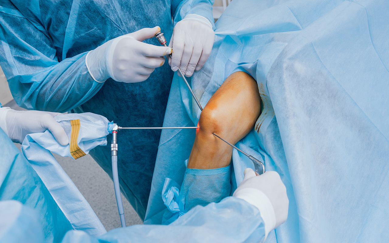
In more than 90% of the cases, a meniscus tear is repaired by arthroscopy in a procedure called debridement.
A debridement is an arthroscopic procedure in which an arthroscope is inserted into the knee in order to repair the torn tissue.
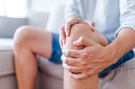
Meniscal tears commonly take place because of accidents or injuries. Patients who have a torn meniscus will experience locking, clicking, swelling, and pain in the knees.
Treatment & Procedures
Meniscus Treatment
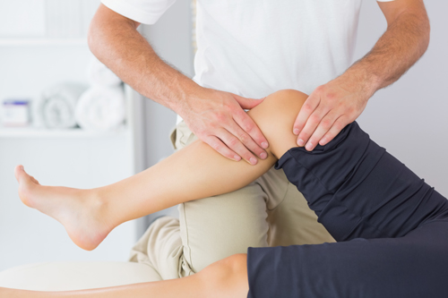
Your doctor will determine if you have a tear by conducting a physical examination and using imaging tests like X-rays and MRI to see the extent of the tear.
Surgical Procedure
- This procedure takes less time to heal than meniscus repair surgery because it’s the least invasive procedure.
- A meniscus debridement is also a better option compared to other surgical procedures because it’s hard to repair a torn meniscus because of the lack of blood supply to the tissue.
- The surgery is performed by making 2-3 puncture wounds in the knee and inserting an arthroscope to remove loose pieces of cartilage stuck between the bones.
Recovery
- The patient can start walking and resume his daily activities in a short period of time after completing a period of physical therapy.
- The patient will typically bear as much weight as they can tolerate but they may need the help of crutches for a few days following the surgery.
- Swelling starts improving after the first week. Patients with desk jobs can return to work within one or two days. More physical laborers who are involved in construction work may take longer to recover.
- Patients typically return to sports or exercise by 4 to 6 weeks following a short period of physical therapy.
Loose Body Removal
What is a Loose Body of Bone Removal?
Loose body removal is an arthroscopic surgery used to remove bits of bone, cartilage, or other tissue stuck between knee joints. It’s a minimal invasive surgery and is performed under a local anesthetic.
Loose bodies are bits of bone, cartilage that float around the body and get caught in joints. These loose bodies cause pain and locking in the joints.
Loose bodies are mostly caused due to intensive physical activity and are common among athletes and jobs where manual labour is common.
Diagnosis
Loose bodies are most commonly diagnosed through physical examination and imaging tests. X-rays, MRI, CT scans, and Arthrography are some of the tests used to determine if someone has loose bodies.
Surgical Procedure
- If the patient has loose bodies locked in their joint, they can’t extend their joints fully. So, it’s important to get them removed as soon as possible.
- Loose bodies can be removed surgically or non-surgically depending on the severity of the condition.
- Arthroscopy is the most preferred surgical method to remove the loose bodies. First, the patient is positioned such that the physician can see the knee clearly.
- Next, the area is cleaned and sterilized. Then, the surgeon makes 2-5 incisions around the front of the knee.
- A camera is inserted from one of the incisions while the arthroscopic tools are inserted from the other incisions.
- The surgeon uses a camera to survey the area and identify the loose bodies. In some cases, the loose bodies may be difficult to find.
- Once the loose bodies are found, the surgeon uses a suction cup or grasping tool to remove the loose bodies in the knees.
- The instruments are then removed and the incisions are sutured and bandaged. The knee will heal within a month.
Cartilage Defects
Treating Cartilage Defects
Cartilage is tissue present between the joints. The function of the cartilage is to cushion the joints, absorb the impact, and support our body weight while we walk, run, or stretch.
Some body parts like ears are made entirely of cartilage. In kids, the end of larger bones are made of cartilage which later transform into bones as we age.
Unlike all other tissue in our body, the cartilage doesn’t have a blood supply to it. So, defects in the cartilage take longer to heal.
There are 3 kinds of cartilage in our body:
Elastic Cartilage: This type of cartilage is very pliable and is commonly found in our ears and nose.
Fibrocartilage: This type of cartilage is very tough and is found between the vertebrae. It’s also found between the hips and pelvis.
Hyaline Cartilage: This type of cartilage is both tough and elastic. It’s present between the joints, around the windpipe and in the ribs.
When the cartilage is damaged it causes pain and inflammation. In some cases it may also lead to a type of disability called articular cartilage.
What Causes a Cartilage Damage?
Patients who suffer from cartilage damage also don’t have a free range of motion as they can’t move their joints fully.
Cartilage damage can also cause the joints to lock and lead to bleeding in the joints. This bleeding is called haemarthrosis.
If a patient has haemarthrosis, there will also be bruising in the joints.
Cartilage Damage is caused due to a direct impact to the joint, lack of movement or wear and tear of the joint due to old age.
What are the Symptoms of a Sprain?
Symptoms of a sprain include:
- Pain
- Swelling
- Bruising
- Hearing a pop noise when the injury takes place
- Difficulty in moving the affected area without experiencing pain.
How is Cartilage Damage Diagnosed?
Cartilage damage is diagnosed using an MRI. Treatment depends on the extent of damage caused to the cartilage.
The doctor might prescribe anti-inflammatories or ask you to undergo a surgery if the damage is too extensive.
Typical Surgical Procedures
Debridement: A portion of the cartilage is trimmed and shaved off so that it doesn’t irritate the other parts of the body.
The procedure is done using small arthroscopic instruments like a mechanical shaver.
Mosaicplasty: Undamaged cartilage is moved to the area where there’s extensive cartilage damage. This is generally only used for patients whose cartilage damage is limited to 2 cm.