Dr. S. Arumugam and his team are sufficiently experienced and equipped to deal with knee injuries and conditions.
The most common knee conditions and their treatments.
- Osteoarthritis of the knee
- Rheumatoid Arthritis
- Meniscal Tear
- ACL Injuries
- PCL Injuries
- Chondral Injuries
- Collateral Ligaments Injuries
- Kneecap Disorders
- Knee Fractures
- Knee Muscle and Tendon Injuries
- Painful Knee Replacement
Osteoarthritis of the Knee
What is Osteoarthritis?
Osteoarthritis (OA) or just arthritis is caused when the soft tissue called cartilage surrounding the bones is worn away. This wearing away causes immense pain because the bones start grinding each other.
Osteoarthritis can affect any joint in the body like the knee joint, hip joint, etc. Osteoarthritis of the knee is more common than other types of arthritis in India.
Who gets affected by Arthritis?
Osteoarthritis is more common in the populations above the age of 45. Men are prone to get arthritis below the ages of 45 while women are more prone to arthritis above the ages of 45.
In India alone, more than 180 million people battle arthritis every day. Some of the common risk factors of arthritis are:
- Old age
- Gender (Women are more prone to get arthritis than men)
- Obesity
- Injured joints
- Repeated stress on the joints.
- Bone deformity
- Genetics
How is Osteoarthritis diagnosed?
Osteoarthritis progresses as time goes by. The most common symptoms for this disorder are:
- extreme joint pain
- swelling around the knee joint
- limited range of motion without experiencing any pain
- Cracking or popping noises in your knee whenever you try to move it.
There are many different ways to diagnose Osteoarthritis
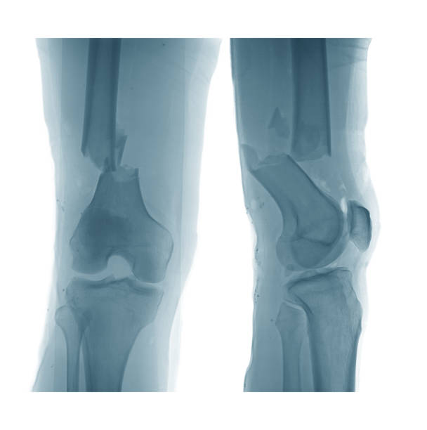
- X-rays. An X-ray is the best indicator of arthritis because tissues don’t show up on it. An arthritic knee will have minimal/ no gap between bones, whereas a healthy knee will have plenty of gap between the two bones.
- MRI scans are used to get a better view of organs and bones in our body. If your doctor suspects that you have arthritis, they will look for damage to the cartilage, tears in ligaments and check if there are any signs of inflammation.
X-rays alone are enough to diagnose arthritis in most cases.
What treatment will I be given for Osteoarthritis?
Firstly, surgery is not the only option to get rid of osteoarthritis. It’s the last resort and Dr. Arumugam will suggest a surgery only when alternate treatment methods like food habits, vitamin tablets and injections don’t lessen the pain.

Rheumatoid Arthritis
What is Rheumatoid Arthritis ?
Rheumatoid Arthritis is a joint disease, and a knee condition caused when the immune system mistakes healthy tissues and joints for invaders and attacks them. It’s an auto-immune disorder, and in severe cases,the organs will also be affected.
Who gets affected by Rheumatoid Arthritis?
Although Rheumatoid Arthritis can affect people of any age, it is most common among 30-50-year-olds. Again, this doesn’t mean that it cannot affect children and elderly patients.
Women are more likely to be affected by Rheumatoid Arthritis than men. Risk factors for Rheumatoid Arthritis include:
- Obesity. Several studies have shown that the risk for Rheumatoid Arthritis increases with the increase in weight.
- Smoking. According to a study, children of parents who smoke are more likely to be affected by Rheumatoid Arthritis.
- Family history. If anyone in your family has Rheumatoid Arthritis, you’re also more likely to be affected by it.
How is Rheumatoid Arthritis diagnosed?
Unlike Osteoarthritis, Rheumatoid Arthritis symptoms start escalating within a short span of time. You’ll start feeling fatigued, be plagued by mild fevers, have morning stiffness along with pain, inflammation, and redness.
Moreover, there are periods in which these symptoms worsen and subside. The former is called having a “flare”.
Rheumatoid Arthritis is difficult to diagnose in the early stages. And if the symptoms mentioned above persist for more than 6 weeks, your doctor might consider these to be symptoms of RA.
You’ll also be asked to take blood tests to confirm if you have Rheumatoid Arthritis.
What treatment will I be given for Rheumatoid Arthritis?
There is no cure for Rheumatoid Arthritis because it’s an auto-immune disorder. But medications and physiotherapy can make your life easier.
You’ll be prescribed certain anti-inflammatory drugs and immuno-suppressants to keep your symptoms from worsening.
Regular exercise and physiotherapy will also help you deal with the inflammation and pain. When everything fails and your pain worsens, surgery is an option.
ACL Injury
What is an ACL injury?
The Anterior Cruciate Ligament (ACL) is a connective tissue that connects the thigh bone and the shin bone. This ligament gets torn when one stops or changes direction suddenly.
Who gets affected by ACL?
Like Meniscal tears, ACL tears are most likely to affect sportspeople. But anyone who stops suddenly or puts all their body weight on the knees may undergo an ACL tear.
This injury is most common in basketball, soccer, and football players. Skiing can also cause this injury.
Women who play sports are seven times more likely to tear their meniscus than men. So far, sports medicine experts haven’t been able to decode the reason behind this.
How is ACL diagnosed?
If you’ve torn your ACL, you’ll:
- Hear a “pop” sound in your knee.
- Feel that your knees are suddenly giving away. The knees will start buckling and won’t be able to support your weight.
- Have a swollen and deformed knee.
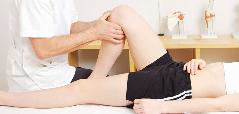
Your doctor will conduct a physical examination and compare your injured knee to the non-injured one. They’ll check your knee and ask you to bend it in order to see how far it can be bent.
If nothing is clear from performing a physical examination, your doctor will ask you to get an MRI or perform an arthroscopy to see if your ligament is torn.
How is the ACL tear treated?
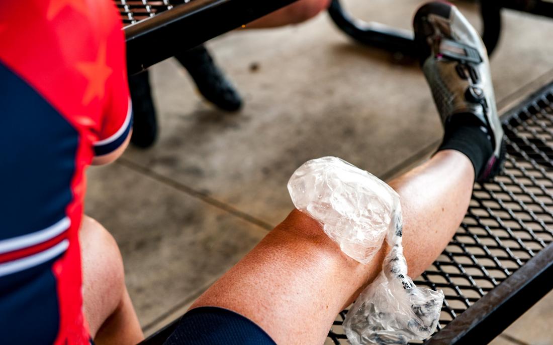
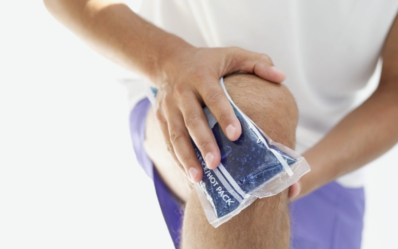
Normally, the ACL tear is treated by the RICE method. You’ll have to Rest, Ice the joint, apply Compression, and Elevate the injured knee.
Your doctor will prescribe non-steroidal anti-inflammatories and ask you to wear knee braces to help you walk around.
Once the pain in your knee subsides, your doctor will ask you to undergo physiotherapy to stabilise and strengthen your knee joints. You’ll be able to walk without feeling as though your knee might give away any moment.
If your ACL tear is severe, you might have to get a surgery. The RICE method is followed even before surgery to reduce some of the swelling.
Once the inflammation and pain have lessened, the Anterior Cruciate Ligament is reconstructed using tissues or tendons from you or from another suitable donor.
PCL Injury
What is a PCL injury?
Posterior Cruciate Ligament is also a connective tissue that connects the bones in the knee. The Anterior Cruciate Ligament (ACL) and Posterior Cruciate Ligament (PCL) form an X-shaped band in the knee to support it.
The PCL prevents the knee from bending backward too much. When the PCL is torn, the front of the knee hurts, and the knee feels “different.”
Who gets affected by PCL?
A Posterior Cruciate Ligament tear takes place during accidents when the knee smashes against the dashboard of the vehicle with force.
It can also take place when a player falls forward on a bent knee with a considerable amount of force. This is common in football and basketball.
Again, these injuries can be had by anyone but are more common in athletes and other sports people.
How is PCL diagnosed?
Some of the first symptoms that you see when you have a Post Cruciate Ligament tear are:
- Mild swelling in the knee joint without a feeling that the knee is about to buckle.
- Moderate pain in the back of the knee that worsens when you bend it.
- Some pain in the front of the knee that worsens when you run or slow down.
When you show up for a visit to the doctor, they’ll ask you to describe the fall to get an idea of what your injury might be.
Then the doctor will conduct a physical examination on your injured knee and compare it to the uninjured knee to see how much the knee has deformed or swollen. They might also tug your knee slightly to know exactly where it hurts.
How should a PCL tear be treated?
No matter how severe your tear is, your first priority must be to reduce the swelling and pain in the joint.
To do this, you’ll have to follow the RICE method. Get plenty of rest, Ice the swollen area, apply Compression, and Elevate the joint.
Once the swelling has reduced, you’ll be asked to undergo therapy or surgery depending on the severity of the tear.
Chondral Injuries
What are Chondral Injuries?
Chondral injuries are tears or scrapes on the cartilage. These scrapes might be caused by the gradual wear and tear of the cartilage caused due to injuries.
Chondral injuries can also be caused when bits and pieces of cartilage break and float around the knee and cause the joints to lock.
Who gets Chondral Injuries?
The elderly population are more prone to getting chondral injuries as a result of falls. More than 10% of people aged over 40 are affected by Chondral injuries.
How are Chondral injuries diagnosed?
Chondral injuries are difficult to diagnose because the symptoms may not be apparent.
A regular physical exam may show swelling but it may not be enough to rule it as a chondral injury. X-rays and MRIs are also not that effective in diagnosing chondral injuries.
The most accurate way to diagnose a chondral injury is by using an arthroscope. The doctor will insert an arthroscope inside the joint and visually assess the damage caused to the joints.
How are chondral Injuries treated?
Chondral injuries can be treated by surgical and non-surgical means. Non-surgical methods include exercise, changes to diet, injections, supplements, and weight management.
Surgical methods include shaving, microfracture abrasion, etc. Choosing a surgical method depends on many factors like:
- Age of the patient.
- Weight of the patient.
- Where the tear is located.
- The size of the tear.
- The patient’s future goals
- How active the patient is and how much they’ll be able to engage in therapy after the operation.
What are the symptoms of Chondral Injuries?
The symptoms of a chondral injury may not be apparent because they may take years to develop.
But the most common symptoms of a chondral injury include:
Swelling: When broken fragments of cartilage float around in the knee, it causes pain and swelling.
Giving way: Your legs might feel as though they’re about to buckle.
Locking: The broken pieces of cartilage can cause your joints to lock, and you won’t be able to move your knees fully. You’ll also hear a clicking noise in your knees.
Collateral Ligament Injuries
What are Collateral Ligament Injuries?
Collateral ligament are present on the left and right side of your knee, and their function is to prevent your knee from moving sideways.
They extend when you extend your knee and relax when your knees are retracted.
Think of them as a patch on either side of your knee that prevents it from moving too much to the left and right.
The Lateral Collateral Ligament (LCL) runs on the outer side of the knee while the Medial Collateral Ligament (MCL) runs on the inner side of the knee.
Collateral Ligament Injuries take place when your knee twists too much towards the side or when you’re hit by a direct blow to the sides of your knee.
Who gets Collateral Ligament Injuries?
Sportspersons are more prone to injuring their Collateral Ligaments while falling. People who play soccer, football, and basketball regularly also have a higher chance of acquiring Collateral Ligament Injuries.

What are the symptoms of Collateral Injuries?
If you’ve collateral injuries you’ll experience the following symptoms.
- A loud pop during the time of the injury.
- Your knee feels like it’s going to give away sideways.
- The knee locks when you try to move.
- Inflammation in the knee.
- The insides and outsides of your knee hurt.
How are Collateral Ligament Injuries diagnosed?
Collateral Injuries can be diagnosed using an X-ray or MRI.
A Magnetic Resonance Imaging (MRI) scan helps detect any stretching or tears in the tissue through 3-D images. So, it’ll help your doctor find out if you’ve a Collateral Ligament Injury.
X-rays only detect broken bones. So, this might not be sufficient for your doctor to make a diagnosis.
How are Collateral Injuries treated?
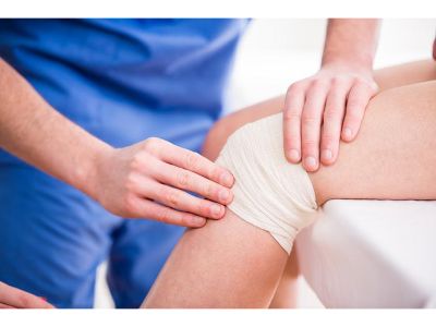
Like other ligament injuries, collateral injuries can be treated with the RICE method. RICE stands for Rest, Ice, Compress, Elevate.
Rest your leg.
Ice the affected area at least 3-4 times a day for 15-20 minutes.
Compress your knee using an elastic wrap or bandage.
Elevate your leg above your heart.
You can ask your doctor for anti-inflammatories and painkillers too. You’ll also have to work with a physiotherapist during this period to regain strength and walk properly
If the pain is unbearable even after self-care, you must consult a doctor.
Kneecap Disorder
What is a Knee Cap Disorder?
The Kneecap or Patella is a bone connecting the thigh bone and the shin bone in the knee joint. It protects the knee joint and connects the thigh bone and shin bone through muscles and ligaments.
When the Kneecap is broken, it shifts outwards or inwards when the knee is bent or straightened.
A Kneecap/Patellar injury can be caused due to:
- Weakening of muscles in the thigh.
- Tightening or loosening of muscles, ligaments, and tendons.
- Strenuous physical activity that involves twisting of the knee.
- Knee injury that dislocates the Kneecap towards the outer side of the leg.
- Improper alignment of knee bones.
Who gets affected by Kneecap Disorder?
Patellar Disorder is more likely to affect sports people who tend to jump on hard surfaces. For example, basketball and volleyball players.
It’s also common in sports where players tend to rotate their torso a lot. For example, baseball.
A patellar injury can also be caused due to an injury or accident as mentioned above.
What are the symptoms of a Patellar/Kneecap Disorder?
- Severe pain in the knee joint while squatting or running.
- A feeling of the knee giving away while you’re walking.
- Popping or grinding in the knee cap whenever you bend or straighten your leg.
You’ll also have severe inflammation if your kneecap disorder is severe. If this happens, consult your doctor.
How is a Patellar/Kneecap Disorder Diagnosed?
Your doctor will ask you questions and conduct a physical examination to determine if you’ve a knee injury.
X-rays only detect broken bones. So, this might not be sufficient for your doctor to make a diagnosis.
He’ll also ask you to get an MRI to better diagnose your injury.
How is Patellar/Kneecap Disorder treated?
To treat a patellar disorder, you must:
- Apply ice to the knee regularly.
- Take a break from physical activity like running and squatting.
- Stretch regularly and focus on doing exercises that improve strength. Speak to a physical therapist to help you choose exercises according to your condition.
- Use the knee brace for extra support
- Put on the right shoes or use shoe inserts (orthotics) to improve the position of your feet.
Knee Fractures
What are Knee Fractures?
The patella is a bone present in the knee that connects thigh bones and shin bones with the help of muscles, ligaments and tendons.
A Knee Fracture is caused when there are cracks in the patella. The cracks in the patella can be mild or even result in a complete breakage of the knee bone.
How does a Knee Fracture occur?
A Knee Fracture occurs when the patella is broken due to falls, sports activities, and direct blows to the knee.
In cases of accidents, there is also a risk of infection if the wound starts bleeding.
What are the symptoms of a Knee Fracture?
The symptoms of a knee fracture include:
- Immediate inflammation, bruising, pain, and tenderness after the injury.
- You can’t touch your knee without extreme pain.
- It’s impossible to bend the knee
- The pain gets worse every time you try to move.
- Bones start grating against each other whenever you try to move your leg.
- Your leg changes shape.
How is Knee Fracture Treated?
- If the knee has an open wound, you might need surgery to treat the fracture. The doctors will also provide treatment to reduce/prevent the infection and control the bleeding.
- The doctor will proceed to move the bones to the right position. You’ll be given the right medication so that you don’t feel any pain during the procedure.
- If the fracture is complicated, your surgeon will also use pins, wires, screws, plates, and rods to hold the bones in place.
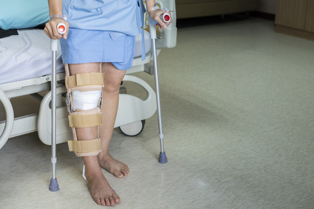
- Your doctor will put your leg in braces, knee immobilizers, or casts to support it and keep it from moving while it heals.
- If you have your leg in a cast, make sure to keep it dry and cover it with plastic. You mustn’t insert anything into it, as it might lead to further infection and injury.
- In case of a mild fracture, apply ice packs to the affected area, and keep your knee as straight as possible.

How is Knee Fracture diagnosed?
Your doctor will ask you questions and conduct a physical examination to determine if you’ve a knee injury.
X-rays only detect broken bones. So, this might not be sufficient for your doctor to make a diagnosis.
He’ll also ask you to get an MRI to better diagnose your injury.
Knee Muscle and Tendon Injuries
What are Knee Tendon Injuries?
Tendons are bands of tissue that connect muscles and the bones together. The Patellar Tendon goes over the patella (kneecap) and works in conjunction with the quadriceps (thigh muscles) to help straighten your leg.
When this tendon becomes swollen, it’s called Tendonitis or Jumper’s Knee because it’s more common among sportspeople who jump often.
The swelling may give way to a Patellar Tendon Tear which is characterised by pain in front of the leg. The tendon in the knee can also get torn because of an accident or injury.
Who is more prone to Knee Tendon Injuries?
Knee Tendon Injuries are more common in people who are involved in sports or activities that involve a lot of running and jumping. For example, basketball and gymnastics.
What are the symptoms of a Patellar Tendon Tear?
Symptoms of a Patellar Tendon Tear include:
- Pain in the front of the knee while you jump or hop
- Inflammation
How is a Patellar Tendon Tear diagnosed?
A Patellar Tendon Tear can be diagnosed in different ways:
- Your doctor might conduct a physical knee examination to determine if you have a patellar tendon tear. They’ll ask you about the activities you’re involved in or if you’ve recently had a blow to your knee.
Patellar Tendon Tear cannot simply be detected using physical examination. So, your doctor might ask you to get an MRI to get a better idea of the injury and the damage caused because of it.
How is a Patellar Tendon Tear treated?
There are different ways to treat a Patellar Tendon Tear:
These include:
- Physiotherapy. Your doctor will prescribe plenty of rest and ask you to work with a physiotherapist to get back on your feet.
- Plasma Injections. These injections will repair the tissue damage and cause your tendons to heal.
- Shockwave Therapy is a safe and effective process to revive the body’s natural healing process.